


线粒体肿胀是一种常见的线粒体增大类型,是由于水(和溶质)进入细胞器,并且可以由许多造成细胞损伤的物质引起。 线粒体肿胀,由粗面内质网的肿胀和水泡组成(另见第 430 页)。 根据过程所达到的阶段,可以看到线粒体形态的各种变化。 线粒体中有两个隔室:(1) 外室或膜间室位于线粒体包膜的两层膜之间,并作为潜在空间或裂缝状或管状空间延伸在嵴内; (2) 包含线粒体基质的内室。
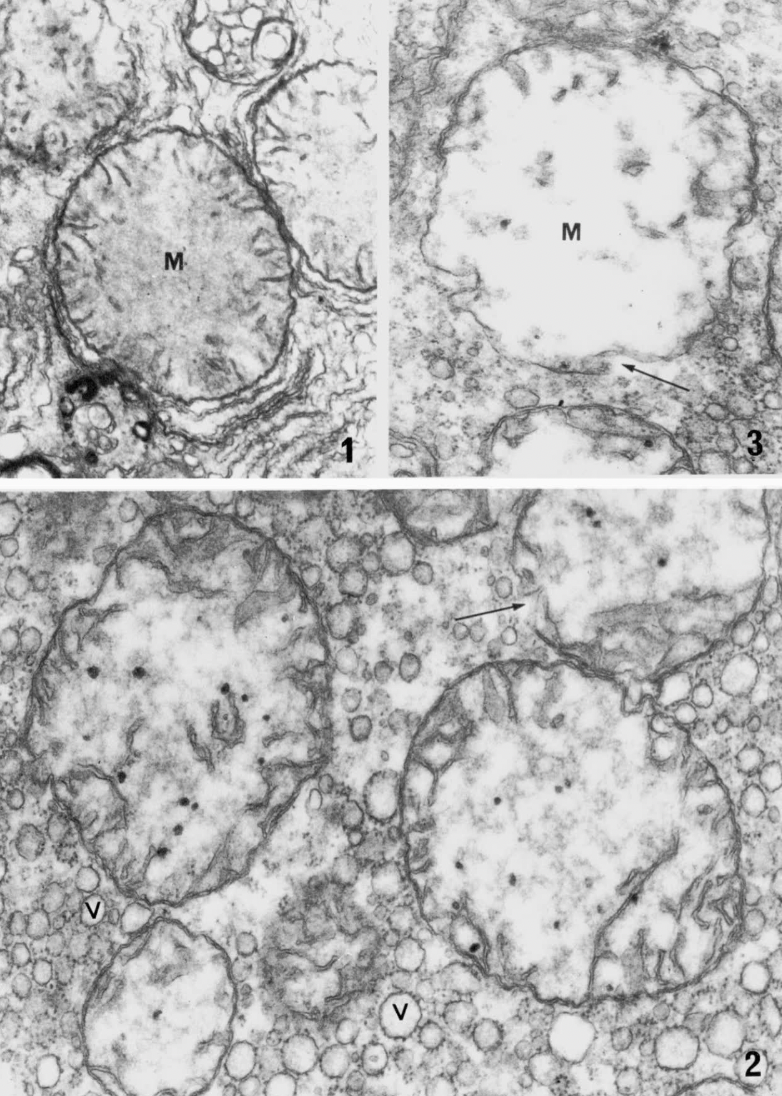
Mitochondria from the liver of a rat bearing a carcinogen-induced subcutaneous sarcoma, showing varying degrees (increasing from Figs. 1 to 3) of swelling of the matrical chamber (Ghadially and Parry, unpublished electron micrographs) Fig. 1. A slightly swollen mitochondrion (M) showing peripherally placed cristae and a fairly homogeneous, medium-density matrix. Intramitochondrial dense granules are absent. X 32 000 Fig. 2. Markedly swollen mitochondria with peripherally placed, disorientated, and disintegrating cristae. The matrix has a patchy appearance and a break (arrow) is evident in the wall of a mitochondrion. Dense granules are abundant. Note also the vesiculation (V) of the rough endoplasmic reticulum. In pathological terms, this cell is showing cloudy swelling. X 46000 Fig. 3. This grossly swollen mitochondrion shows frank cavitation of the mitochondrial matrix (M) and loss of cristae. The lower half of the mitochondrial wall is attenuated and probably ruptured (arrow). X 46000
来自患有致癌物诱导的大鼠肝脏的线粒体,显示不同程度(从图 1 到图 3 增加)的基质室肿胀
图 1. 轻微肿胀的线粒体 (M) 显示外围放置的嵴和相当均匀的中等密度基质。 不存在线粒体内致密颗粒。 X 32 000
图 2. 线粒体明显肿胀,嵴位于外围。 基质呈斑驳状,线粒体壁上有明显的断裂(箭头)。 致密颗粒丰富。 有粗面内质网的小泡 (V)。 在病理学上,这个细胞呈浑浊肿胀。 X 46000
图 3. 这种严重肿胀的线粒体显示线粒体基质 (M) 明显空化和嵴缺失。 线粒体壁的下半部分可能破裂(箭头)。 X 46000
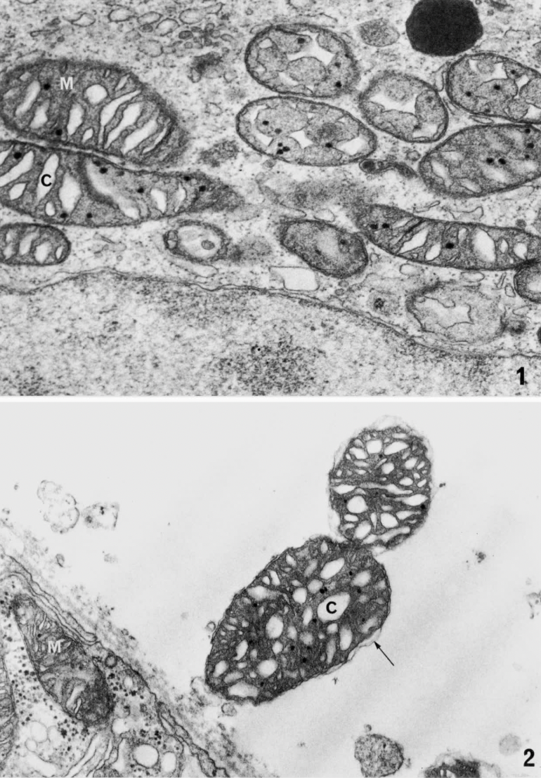
Fig. I A monkey kidney cell from a culture infected with herpesvirus. Mitochondria cut in various planes clearly show markedly ballooned cristae (C) and a moderately dense matrix (M), but there is no separation of the outer and inner mitochondrial membranes. This (and also Fig. 2) is the appearance which is sometimes referred to as a ' condensed configuration'. Several matrical dense granules are present. X 41 000 Fig. 2. Two mitochondria, which no doubt escaped while the tissue (frog kidney) was being minced in osmium, are now seen lying free in the embedding medium. Both show markedly ballooned cristae (C), a dense matrix and some intramitochondrial dense granules. There is focal separation of inner and outer membranes (arrow) in one mitochondrion, while in the other the separation is much more extensive. These mitochondria are grossly swollen compared to the mitochondria (M) within the kidney tubule. X 32 000
图 1 来自感染了疱疹病毒的猴肾细胞。 在不同平面上切割的线粒体清楚地显示出明显膨胀的嵴 (C) 和中等密度的基质 (M),但线粒体内外膜没有分离。 X 41 000
图 2. 两个线粒体。 两者均显示明显膨胀的嵴 (C)、致密基质和一些线粒体内致密颗粒。 一个线粒体中的内膜和外膜(箭头)存在分离,而在另一个线粒体中则更为广泛。 与肾小管内的线粒体 (M) 相比,这些线粒体严重肿胀。 X 32 000
在肥大的线粒体中,嵴的密度可能增加,但在肿胀的线粒体中,嵴丢失或移到外围。 在心脏、子宫和骨骼肌中观察到线粒体肥大或数量增加的例子,这些肌肉受到各种病理和实验状态的影响,对组织的功能需求增加。 因此,怀孕期间子宫肌层的显着肥大不仅与线粒体数量和大小的大幅增加相匹配,而且与嵴数量的增加相匹配(Dessouky,1968)。 在急性和力竭运动后的心肌中也观察到类似的线粒体变化(Laguens 等人,1966 年;Pelosi 和 Agliati,1968 年)以及自然疾病或实验情况引起的心脏肥大(Meerson 等人,1964 年;McCallister 和 Brown,1965;Bishop 和 Cole,1969;Zak 和 Rabinowitz,1973)。

An oncocyte from a bronchial mucosal gland of man, showing cytoplasm packed with mitochondria. X 24 000
来自人类支气管粘膜腺的癌细胞,显示细胞质中充满了线粒体。 X 24 000