


An optimized protocol for immuno-electron microscopy of endogenous LC3
PMID: 35387562 PMCID: PMC9673964 DOI: 10.1080/15548627.2022.2056864
MAP1LC3/LC3(微管相关蛋白1轻链3)被广泛用作不同成熟阶段自噬细胞器的标记物。电子显微镜(EM)结合免疫标记是唯一能够揭示LC3标记细胞器超微结构特征的技术。然而,免疫EM的内源性LC3蛋白已被证明是困难的。在这里,我们测试了一系列商业可获得的抗体,并应用不同的标记条件,提出了一种优化的LC3免疫EM程序。我们使用超薄冷冻切片和蛋白A-胶体金或金增强标记,将内源性LC3定位于饥饿细胞或组织中,在质子泵抑制剂bafilomycin A1存在或不存在的情况下。我们将LC3定位于早期和晚期自噬细胞器,可以通过其形态分类。通过切片相关光电子显微镜(CLEM),我们展示了相似的荧光LC3阳性标记可以代表不同的自噬中间体。我们还展示了我们的方法足够强大,可以同时标记内源性LC3和其他溶酶体和自噬标记物,如LAMP1或SQSTM1/p62,并且可以用于定量方法。因此,我们证明了从2.5小时到24小时的bafilomycin A1处理不会抑制自噬体与溶酶体的融合,但会导致LC3阳性物质在自溶体内积累。总之,这是第一项在超微结构分辨率下呈现内源性LC3定位的广泛概述研究,无需细胞渗透化,并使用商业可获得的抗体。
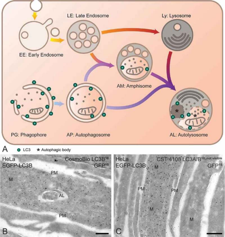
Cosmo Bio anti-LC3B specifically recognizes LC3B in EGFP-LC3B transfected cells. (A) Schematic overview of endo-lysosomal and autophagic compartments, outlining their characteristic morphological features and presence of LC3. (B-C) EGFP-LC3B transfected cells were fixed with 2% PFA+0.2% GA for 3 h and double labeled for LC3B and GFP and protein A gold (PAG). (B) The labeling intensities of anti-GFP (1:400; PAG15) and Cosmo Bio anti-LC3B (1:10; PAG10) correlate in cells with high and low EGFP-LC3B overexpression. (C) Example of double labeling with anti-GFP (1:400; PAG15) and another LC3 antibody (CST, 4108, 1:15; PAG10) showing only PAG15 on EGFP-LC3B overexpressing cells. AL, autolysosome; M, mitochondrion; PM, plasma membrane. Scale bars: 300 nm.
Cosmo Bio的抗LC3B能特异性识别EGFP-LC3B转染细胞中的LC3B。(A)内源性溶酶体和自噬细胞器的示意图,概述其特征形态特征和LC3的存在。(B-C)EGFP-LC3B转染细胞固定于2% PFA+0.2% GA中3小时,并进行LC3B、GFP和蛋白A金(PAG)的双重标记。(B)抗GFP(1:400;PAG15)和Cosmo Bio抗LC3B(1:10;PAG10)在EGFP-LC3B高和低表达细胞中的标记强度呈正相关。(C)双重标记示例,使用抗GFP(1:400;PAG15)和另一种LC3抗体(CST,4108,1:15;PAG10),仅在过度表达EGFP-LC3B的细胞上显示PAG15。AL,自溶体;M,线粒体;PM,细胞质膜。标尺300纳米。
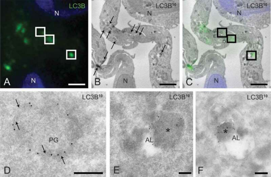
Correlative light electron microscopy (CLEM) of endogenous LC3B on ultrathin cryosections. (A) Ultrathin (60–70 nm) cryosection of starved, BafA1-treated U2OS cells (fixation 4% PFA ON) labeled for LC3B (1:10), rabbit anti-mouse IgG, Alexa Fluor 488-conjugated donkey anti-rabbit IgG and PAG10, showing IF puncta (green) and nuclei (Hoechst, blue). (B) Low magnification EM picture of same section as in (A). Arrows point to LC3B-immunogold labeled compartments (gold not visible at this magnification). (C) Overlay of the fluorescent and EM images in (A) and (B). Higher magnifications of the boxed areas in (C) are shown in (D-F). (D) A phagophore (PG) is visible as a ring of LC3B-positive vesicles. Arrows indicate visible membrane contours. (E, F) Two examples of autolysosomes (AL). LC3B (PAG10) is predominantly associated with autophagic content located in the AL lumen (indicated by asterisks). Note that the higher magnifications in D-F are rotated relative to B. N, nucleus. Scale bars: 2 µm (A-C), 200 nm (D-F).
在超薄冷冻切片上的内源性LC3B的相关光电子显微镜(CLEM)。(A经BafA1处理的U2OS细胞的超薄(60-70纳米)冷冻切片(固定4% PFA ON),标记LC3B(1:10)、兔抗小鼠IgG、Alexa Fluor 488标记的山羊抗兔IgG和PAG10,显示了IF puncta(绿色)和细胞核(Hoechst,蓝色)。 (B)与(A)中相同切片的低放大率电子显微镜图像。箭头指向LC3B免疫金标记的细胞器(在此放大下金颗粒不可见)。 (C)(A)和(B)中荧光和电子显微图像的叠加。 (D-F)(C)中方框区域的较高放大率显示在(D-F)中。 (D)一个嗜吞噬体(PG)可见为LC3B阳性囊泡的环。箭头指示可见的膜轮廓。 (E,F)两个自溶体(AL)的示例。 LC3B(PAG10)主要与位于AL腔内的自噬内容相关联(由星号表示)。请注意,D-F中的较高放大率相对于B是旋转的。N,细胞核。标尺:2 µm(A-C),200 nm(D-F)。
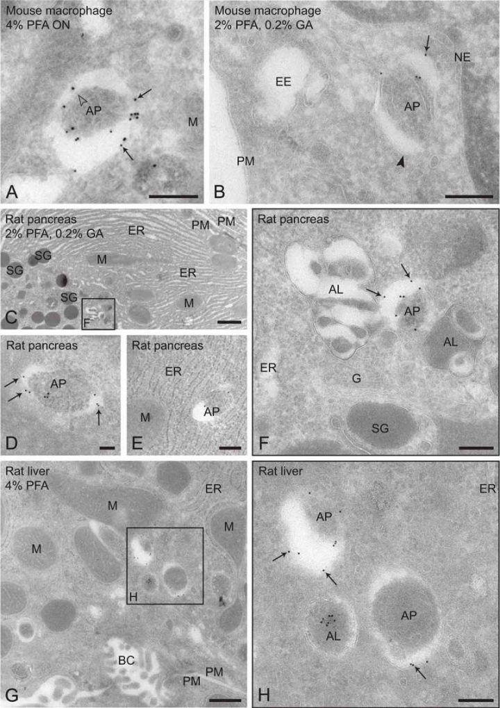
Immuno-EM of endogenous LC3B in primary cells and tissues. (A-B) Primary mouse macrophages were starved for 30 min without addition of BafA1. LC3B labeling (1:6; PAG10, arrows) is detected on autophagosomes (AP) and autolysosomes (AL). Gold particles are associated with the inner (open arrowheads) and outer (black arrowheads) autophagosome membrane. (A) Cells fixed ON with 4% FPA. (B) Cells fixed with 2% PFA+0.2% GA for 2 h. (C-H) In rat pancreas and liver (1:10; PAG10), in the absence of lysosomal inhibitors, LC3B labeling is detected mainly on autophagosomes (AP) and occasionally on an autolysosome (AL). Gold particles are associated with the inner and outer (arrows) autophagosome membrane. (C-F) Rat exocrine pancreas perfusion fixed with 2% PFA+0.2% GA. (C) Overview. (D, E) Examples of autophagosomes (AP) encapsulating mainly ER membranes. (F) Group of 2 (not-labeled) autolysosomes (AL) and an autophagosome (AP) near the Golgi (G), enlarged from the box in (C). LC3 immunogold label is present on inner and outer (arrows) AP membrane. (G, H) Rat liver perfusion fixed with 4% PFA. (H) LC3 immunogold label on a group of autophagosomes (AP) and an autolysosome (AL) in a hepatocyte, enlarged from the box in (G). Arrows indicate LC3 gold on the outer AP membrane. BC, bile canaliculus; EE, early endosome; ER, endoplasmic reticulum; M, mitochondrion; NE, nuclear envelope; PM, plasma membrane; SG, secretory granule. Scale bars: 100 nm (D), 200 nm (A, B, E, F, H), 1 μm (C), 500 nm (G).
原代小鼠巨噬细胞内源性LC3B的免疫电镜。 (A-B)未添加BafA1的情况下,原代小鼠巨噬细胞在饥饿30分钟。 LC3B标记(1:6;PAG10,箭头)检测到自噬体(AP)和自溶体(AL)。金颗粒与自噬体内(空心箭头)和外(黑色箭头)膜相关联。 (A)细胞使用4% FPA固定ON。 (B)细胞使用2% PFA+0.2% GA固定2小时。 (C-H)在大鼠胰腺和肝脏中(1:10;PAG10),在没有溶酶体抑制剂的情况下,LC3B标记主要检测到自噬体(AP)和偶尔的自溶体(AL)。金颗粒与内外(箭头)自噬体膜相关联。 (C-F)使用2% PFA+0.2% GA灌注固定的大鼠外分泌胰腺。 (C)总览。 (D,E)主要包含内质网膜的自噬体(AP)的示例。 (F)一组2个(未标记)自溶体(AL)和一个自噬体(AP)靠近高尔基体(G)的显微图像,从(C)中的方框中放大。 LC3免疫金标记存在于内外(箭头)AP膜上。 (G,H)使用4% FPA灌注固定的大鼠肝脏。 (H)在肝细胞中的一组自噬体(AP)和自溶体(AL)上的LC3免疫金标记,从(G)中的方框中放大。箭头指示外部AP膜上的LC3金。BC,胆小管;EE,早期内体;ER,内质网;M,线粒体;NE,核膜;PM,细胞质膜;SG,分泌颗粒。标尺:100 nm(D),200 nm(A,B,E,F,H),1 μm(C),500 nm(G)。