


Endoplasmic reticulum–associated degradation regulates mitochondrial dynamics in brown adipocytes
内质网 (ER) 在称为线粒体相关膜 (MAM) 的专门 ER 结构域与线粒体结合。 在这里,我们使用三维高分辨率成像来研究缺乏 ER 相关蛋白降解 (ERAD) 的 Sel1L-Hrd1 蛋白复合物的棕色脂肪细胞中具有改变的 MAM 的多形性“巨线粒体”的形成。 棕色脂肪细胞中存在 ERAD 缺陷的小鼠对冷敏感并表现出线粒体功能障碍。 ERAD 缺陷至少部分通过调节 MAM 蛋白 Sigma 受体 1 (SigmaR1) 的周转来影响 ER 线粒体接触和线粒体动力学。 因此,我们的研究提供了对 ER-线粒体串扰的分子见解,并扩展了我们对 Sel1L-Hrd1 ERAD 生理重要性的理解。
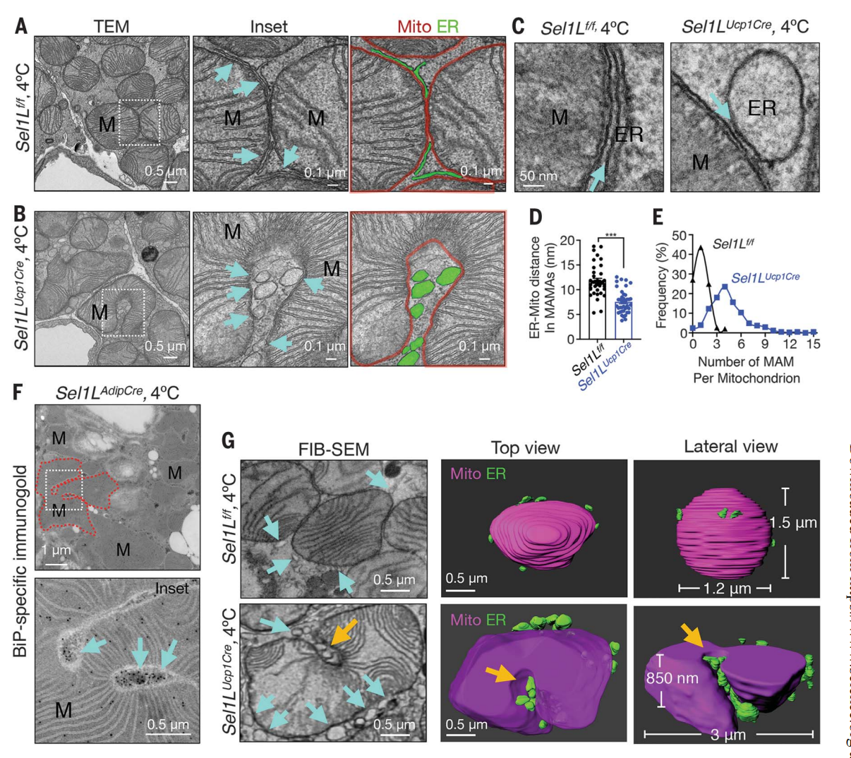
Fig. Sel1L controls ER-mitochondria contacts in cold-stimulated brown adipocytes. (A and B) Representative TEM images of BAT from Sel1Lf/f (A) and Sel1LUcp1Cre (B) mice at 4°C for 6 hours. M, mitochondrion; cyan arrows, MAMs; red lines, mitochondrial membranes; green, ER. The area shown by white dotted lines is enlarged in the inset. (C to E) Representative TEM images of the MAMs in BAT from Sel1Lf/f and Sel1LUcp1Cre mice at 4°C for 6 hours, with quantitation of ER-mitochondrion distance (D) and abundance of MAM per mitochondrion (E). n = 35 and 40 MAMs (D) and n = 450 and 572 mitochondria (E) for Sel1Lf/f and Sel1LUcp1Cre. (F) Representative TEM images of BiP-specific immunogold labeling in BAT from Sel1LAdipCre mice at 4°C for 6 hours. Red dotted line outlines one megamitochondrion; cyan arrows, BiP-positive ER tubule(s). (G) Representative FIB-SEM (left) and the 3D tomography images (300 and 170 slices, 5 nm/slice) of mitochondria in BAT from Sel1Lf/f and Sel1LUcp1Cre mice at 4°C for 6 hours. Magenta, mitochondria; green, ER. Cyan arrows, MAMs; orange arrows, MAMs going through the concave surface of a mitochondrion. All experiments were repeated two to three times. Data are mean ± SEM. ***p < 0.001 by Student’s t test.
图. Sel1L 控制冷刺激棕色脂肪细胞中的 ER 线粒体接触。 (A 和 B) Sel1Lf/f (A) 和 Sel1LUcp1Cre (B) 小鼠的 BAT 在 4°C 下 6 小时的代表性 TEM 图像。 M,线粒体; 青色箭头,MAM; 红线,线粒体膜; 绿色,ER。 插图中白色虚线所示的区域被放大。 (C 至 E) Sel1Lf/f 和 Sel1LUcp1Cre 小鼠 BAT 中 MAM 在 4°C 下 6 小时的代表性 TEM 图像,并对 ER-线粒体距离 (D) 和每个线粒体 MAM 丰度进行定量 (E)。 对于 Sel1Lf/f 和 Sel1LUcp1Cre,n = 35 和 40 个 MAM (D),n = 450 和 572 个线粒体 (E)。 (F) Sel1LAdipCre 小鼠 BAT 中 BiP 特异性免疫金标记在 4°C 下 6 小时的代表性 TEM 图像。 红色虚线勾勒出一个巨线粒体; 青色箭头,BiP 阳性 ER 小管。 (G) Sel1Lf/f 和 Sel1LUcp1Cre 小鼠 BAT 中线粒体的代表性 FIB-SEM(左)和 3D 断层扫描图像(300 和 170 切片,5 nm/切片),在 4°C 下 6 小时。 洋红色,线粒体; 绿色,ER。 青色箭头,MAM; 橙色箭头,MAM 穿过线粒体的凹面。 所有实验重复两到三次。 数据为平均值±SEM。 ***p < 0.001(通过学生 t 检验)。
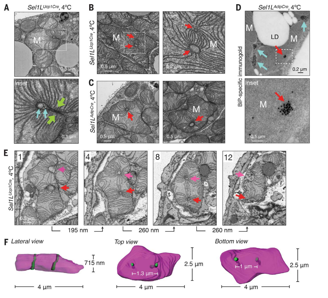
Fig.Sel1L deficiency leads to the formation of megamitochondria with perforating ER tubules. (A) Representative TEM images of BAT in Sel1LUcp1Cre mice housed at 4°C for 6 hours, showing a megamitochondrion wrapping around the tubular structures (cyan arrows). Green arrows, two opposite sides of a mitochondrion. (B and C) Representative TEM images of BAT from Sel1LUcp1Cre (B) and Sel1LAdipCre (C) mice at 4°C for 6 hours, showing megamitochondria with tubular structures (red arrows). (D) Representative BiP-immunogold TEM images of BAT in Sel1LAdipCre mice at 4°C for 6 hours. Red and cyan arrows, mitochondria-perforating ER tubules and perimitochondria ER tubules. (E and F) Representative SBF-SEM images (E) and 3D reconstruction (F) in BAT of Sel1LUcp1Cre mice at 4°C for 6 hours, showing four different slices of a megamitochondrion with two parallel perforating ER tubules (red and magenta arrows). All 12 slices (65 nm/slice) are shown in fig. S8. All experiments were repeated two to three times.
图 . Sel1L 缺陷导致具有穿孔 ER 小管的巨线粒体的形成。 (A) 在 4°C 下饲养 6 小时的 Sel1LUcp1Cre 小鼠中 BAT 的代表性 TEM 图像,显示巨型线粒体包裹在管状结构周围(青色箭头)。 绿色箭头,线粒体的两侧。 (B 和 C) Sel1LUcp1Cre (B) 和 Sel1LAdipCre (C) 小鼠的 BAT 在 4°C 下 6 小时的代表性 TEM 图像,显示具有管状结构的巨线粒体(红色箭头)。 (D) Sel1LAdipCre 小鼠中 BAT 在 4°C 下 6 小时的代表性 BiP-免疫金 TEM 图像。 红色和青色箭头、线粒体穿孔 ER 小管和线粒体周围 ER 小管。 (E 和 F)代表性 SBF-SEM 图像(E)和 Sel1LUcp1Cre 小鼠 BAT 中 4°C 6 小时的 3D 重建(F),显示具有两个平行穿孔 ER 小管的巨线粒体的四个不同切片(红色和洋红色箭头) )。 所有 12 个切片(65 nm/切片)如图 2 所示。 S8。 所有实验重复两到三次。