


Tg1.4HBV-s-rec mice, a crossbred hepatitis B virus-transgenic model, develop mild hepatitis
Scientific Reports volume13, Article number: 22829 (2023)
乙型肝炎病毒(HBV)转基因小鼠表现出良好的先天免疫功能,因此是考虑HBV病理生理学中内在或基于细胞的机制的理想模型。一种高度复制的、鲜为人知的模型是Tg1.4HBV-s-rec株系,它是由积累(Alb/HBs,Tg[Alb1-HBV]Bri44)或缺乏(Tg1.4HBV-s-mut)乙型肝炎表面抗原(HBsAg)的HBV转基因小鼠模型杂交而来。Tg1.4HBV-s-rec肝细胞分泌HBsAg、乙型肝炎病毒外细胞抗原(HBeAg)并产生HBV病毒颗粒。透射电子显微镜观察到病毒颗粒(Tg1.4HBV-s-rec)、核囊形成(Tg1.4HBV-s-mut和Tg1.4HBV-s-rec)以及内质网异常形态(Alb/HBs)。Tg1.4HBV-s-rec和Tg1.4HBV-s-mut中的病毒复制在HBsAg表达和有趣的是HBV核心抗原(HBcAg)和HBV × 蛋白的分布方面存在差异。在Tg1.4HBV-s-mut肝细胞中,HBcAg位于细胞质中,而在Tg1.4HBV-s-rec肝细胞中,HBcAg出现在细胞核中,表明更为有效的复制。最后,Tg1.4HBV-s-rec小鼠表现出轻度肝炎症状,肝功能降低,血清转氨酶升高,这似乎与自然杀伤T细胞的激活有关。总的来说,对Alb/HBs、Tg1.4HBV-s-mut及其F1后代的研究为阐明HBV病理生理学提供了强大的工具,特别是在慢性感染和慢性肝炎的早期HBeAg阳性阶段。
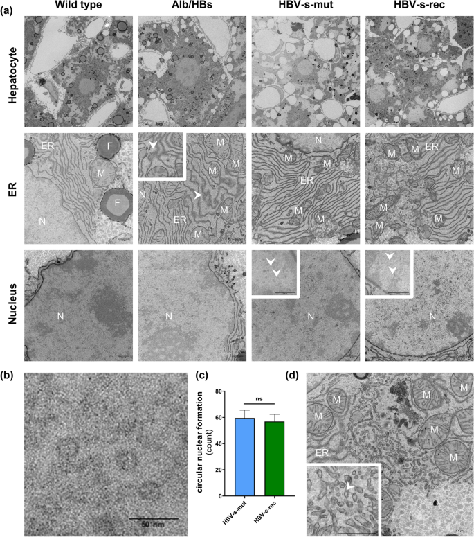
Viral particles are produced in the HBV-s-rec mouse strain. (a) Liver tissue from 6-month-old wild type and HBV-transgenic mice were fixated and contrasted according to a modified OTO protocol. Images are representatives of each mouse strain (group sizes n = 3). General cell morphology of hepatocytes, the endoplasmic reticulum (ER), cytoplasm, and nuclei are visualised using transmission electron microscopy (TEM). (b) Representative high-resolution image of circular structures in a hepatocyte nucleus (HBV-s-mut). (c) Quantification of circular nuclear formations in HBV-s-mut and HBV-s-rec (mean ± SD, counts per nucleus, n = 10). (d) TEM imaging of the Golgi apparatus visualising HBV particle in an HBV-s-rec hepatocyte. M, mitochondria; N, nucleus; F, fat droplets; scale bars, 0.5 µm (a, d) 50 nm (b); nm, nanometer; ns, not significant.
病毒颗粒在HBV-s-rec小鼠株系中产生。(a)采用修改后的OTO方案,固定并对比了6个月大的野生型和HBV转基因小鼠的肝组织。图像是每个小鼠株系的代表性图像(组大小n = 3)。使用透射电子显微镜(TEM)可视化了肝细胞、内质网(ER)、细胞质和细胞核的一般细胞形态学。(b)肝细胞核中圆形结构的代表性高分辨率图像(HBV-s-mut)。 (c)HBV-s-mut和HBV-s-rec中圆形核结构的计量(平均值±SD,每个细胞核的计数,n = 10)。 (d)使用TEM成像可视化了HBV-s-rec肝细胞中的高尔基体,显示HBV颗粒。 M,线粒体;N,细胞核;F,脂滴;比例尺,0.5 µm(a,d)50 nm(b);nm,纳米;ns,不显著。
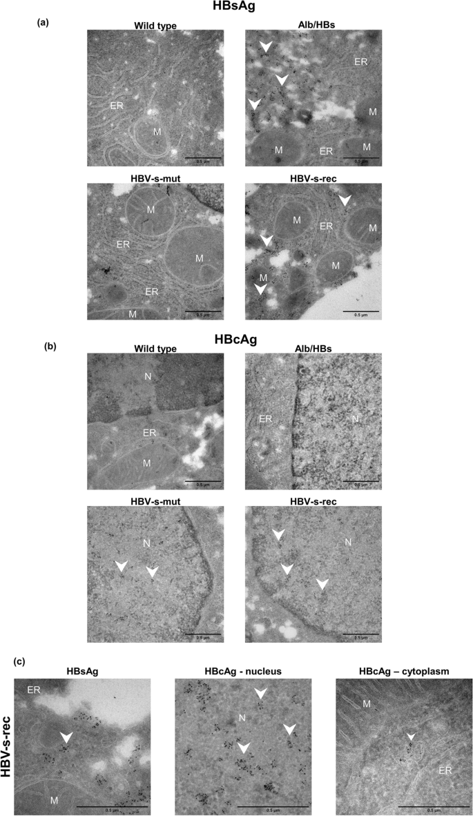
Visualising viral particles in the Tg1.4HBV-s-rec mice. (a) Liver tissue from 6-month-old wild type and HBV-transgenic mice (Alb/HBs, HBV-s-mut, HBV-s-rec) were fixated and contrasted according to an immunogold staining protocol. Images are representatives of each mouse strain (group sizes n = 3). Endoplasmic reticulum area and nuclei were visualised using transmission electron microscopy. (a) Immunogold staining was performed in wild type and HBV-transgenic mouse strains to visualise (a) HBsAg and (b) HBcAg. (c) Representative high-resolution image of viral particle in HBV-s-rec hepatocytes stained for HBsAg and HBcAg. M, mitochondria; N, nucleus; ER, endoplasmic reticulum; scale bars, 0.5 µm.
在Tg1.4HBV-s-rec小鼠中可视化病毒颗粒。(a)采用免疫胶体金方案,对6个月大的野生型和HBV转基因小鼠(Alb/HBs,HBV-s-mut,HBV-s-rec)的肝组织进行了固定和对比。图像是每个小鼠株系的代表性图像(组大小n = 3)。使用透射电子显微镜可视化内质网区域和细胞核。(a)在野生型和HBV转基因小鼠株系中进行了免疫金染色,以可视化(a)HBsAg和(b)HBcAg。(c)HBV-s-rec肝细胞中经过HBsAg和HBcAg染色的病毒颗粒的代表性高分辨率图像。 M,线粒体;N,细胞核;ER,内质网;比例尺,0.5 µm。
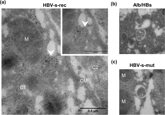
Visualising multivesicular bodies in the Tg1.4HBV-s-rec mice. Liver tissue from 6-month-old HBV-transgenic mice (Alb/HBs, HBV-s-mut, HBV-s-rec) were fixated and contrasted according to an immunogold staining protocol. Images are representatives of each mouse strain (group sizes n = 3). Multivesicular bodies were visualised using transmission electron microscopy. Immunogold staining was performed for HBsAg in HBV-s-rec (a) and Alb/HBs (b) and for HBcAg in HBV-s-mut (c) liver tissue. M, mitochondria; C1, cell 1; C2 cell two; CM, cell membrane.
在Tg1.4HBV-s-rec小鼠中可视化多囊体。对6个月大的HBV转基因小鼠(Alb/HBs,HBV-s-mut,HBV-s-rec)的肝组织进行了固定和对比,使用免疫电镜方法。图像是每个小鼠株系的代表性图像(组大小n = 3)。使用透射电子显微镜可视化多囊体。在HBV-s-rec小鼠的肝组织中进行了HBsAg的免疫金染色(a),在Alb/HBs小鼠的肝组织中进行了HBsAg的免疫金染色(b),在HBV-s-mut小鼠的肝组织中进行了HBcAg的免疫金染色(c)。M,线粒体;C1,细胞1;C2,细胞2;CM,细胞膜。