


In vivo topical gene therapy for recessive dystrophic epidermolysis bullosa: a phase 1 and 2 trial
Nature Medicine volume 28, pages780–788 (2022)
隐性营养不良性大疱性表皮松解症 (RDEB) 是一种终生遗传性皮肤病,与 COL7A1 突变引起的水疱、受伤和疤痕相关,COL7A1 是编码锚定原纤维成分 VII 型胶原蛋白 (C7) 的基因。 在这里,我们评估了 beremagene geperpavec (B-VEC),一种工程化的、非复制的 COL7A1,含有 1 型单纯疱疹病毒 (HSV-1) 载体,用于治疗 RDEB 皮肤。 B-VEC 可恢复 RDEB 角质形成细胞、成纤维细胞、RDEB 小鼠和人 RDEB 异种移植物中的 C7 表达。 随后,一项随机安慰剂对照 1 期和 2 期临床试验 (NCT03536143) 评估了 9 名 RDEB 患者在 12 周内反复接受局部 B-VEC 或安慰剂的匹配伤口。 未发现 2 级或以上 B-VEC 相关不良事件或载体脱落或组织结合皮肤免疫反应物。 HSV-1 和 C7 抗体有时会在基线时出现,或在 B-VEC 治疗后增加,但对安全性或疗效没有明显影响。 满足了 C7 表达、锚定原纤维组装、伤口表面积减少、伤口闭合持续时间以及 B-VEC 治疗后伤口闭合时间的主要和次要目标。 鉴于治疗的伤口比例较小,未评估患者报告的疼痛严重程度次要结果。 由于其他终点的冗余,没有追求全球评估次要终点。 这些研究表明,B-VEC 是一种易于给药、安全耐受的局部分子矫正疗法,可促进 RDEB 患者伤口愈合。
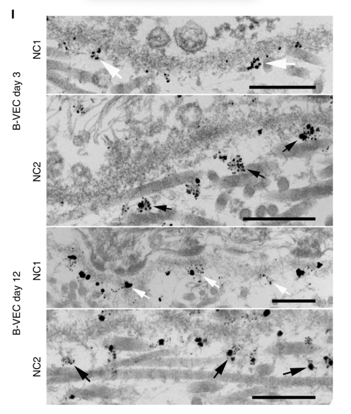
l, Human RDEB skin xenografts were treated with topical B-VEC and analyzed after 3 and 12 days using immunoelectron microscopy with C7 NC1-directed antibodies (NP185), followed by gold nanoparticles (first, third rows; white arrows), and NC2-directed primary antibodies (LH24), followed by gold nanoparticles (second, fourth rows; black arrows). Error bars on all panels represent standard error of the mean of all replicates. Representative of eight grafts treated with B-VEC and two grafts treated with placebo. Scale bars, 300 nm. Data plots, including error bars and P values, were generated using GraphPad Prism v8.3.0.
l,用局部 B-VEC 处理人 RDEB 皮肤异种移植物,并在 3 天和 12 天后使用免疫电子显微镜使用 C7 NC1 定向抗体 (NP185) 进行分析,然后是金纳米粒子(第一行、第三行;白色箭头)和 NC2- 定向一抗 (LH24),然后是金纳米粒子(第二、第四行;黑色箭头)。 所有面板上的误差条代表所有重复的平均值的标准误差。 代表用 B-VEC 治疗的八个移植物和用安慰剂治疗的两个移植物。 比例尺,300nm。 使用 GraphPad Prism v8.3.0 生成数据图,包括误差线和 P 值。
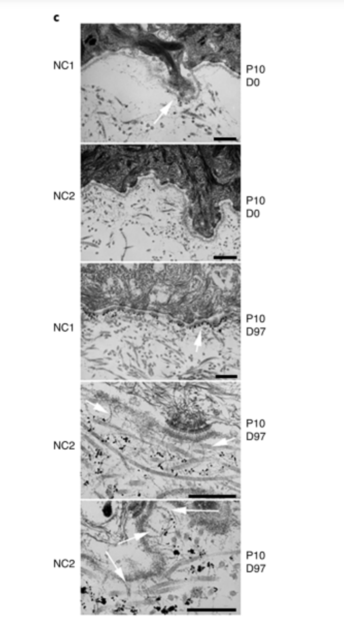
c, Immunoelectron microscopy29 of C7 NC1 and NC2 expression and AFs in B-VEC-treated patient skin. Representative images are shown from patient 10 (collected at the indicated times) and were analyzed with immunoelectron microscopy using antibodies to the C7 NC1 domain (NP185) and C7 NC2 domain (LH24). (C7 NC1 and NC2 expression was assessed in patients whose specimens were amenable to immunoelectron microscopy analysis (n = 3; Supplementary Table 4.) The arrow in the upper panel and center panel shows positive immuno-gold staining for the NC1 domain in the lamina densa region. Arrows in the lower two panels show the presence of mature banded AFs associated with immuno-gold staining for the NC2 domain approximately 300 nm from the lamina densa. Scale bars, 500 nm.
c,B-VEC 处理的患者皮肤中 C7 NC1 和 NC2 表达以及 AF 的免疫电子显微镜检查29。 显示来自患者 10 的代表性图像(在指定时间收集),并使用 C7 NC1 结构域 (NP185) 和 C7 NC2 结构域 (LH24) 的抗体通过免疫电子显微镜进行分析。 (在其标本适合免疫电子显微镜分析的患者中评估了 C7 NC1 和 NC2 表达(n = 3;补充表 4。)上图和中图的箭头显示椎板中 NC1 结构域的免疫金染色阳性 致密区。下面两幅图中的箭头显示存在与距致密层约 300 nm 的 NC2 结构域的免疫金染色相关的成熟带状 AF。比例尺,500 nm。